


How PET CT Scans Can Help Diagnose, Monitor And Treat Cancer
A PET/CT scan is an advanced hybrid imaging technique widely used in cancer diagnosis, staging, treatment planning, and recurrence detection. A PET/CT scan is a whole-body imaging test that combines metabolic and structural imaging to detect, stage, and monitor cancer accurately.
What is a PET/CT scan?
PET/CT stands for Positron Emission Tomography / Computed Tomography scan.
It merges two imaging technologies:
- PET scan (Positron Emission Tomography): A nuclear medicine imaging technique that uses radioactive tracers (commonly FDG – fluorodeoxyglucose) to detect metabolic activity in tissues. Cancer cells typically show higher metabolic activity.
- CT scan (Computed Tomography): Uses X-rays to create detailed cross-sectional images of organs, tissues, and bones.
When combined, PET/CT provides precise anatomical location along with metabolic activity, making it highly effective for detecting tumors, evaluating lymph nodes, and identifying distant metastasis.
What Are Some Common Uses of the Procedure?
A PET/CT scan plays a critical role in modern oncology. Common clinical uses include:
1. Cancer Detection
- Identifies abnormal metabolic activity suggestive of malignant tumors.
- Detects cancers that may not be visible on MRI or CT alone.
2. Cancer Staging
- Determines tumor size and spread (metastasis).
- Evaluates lymph node involvement.
- Helps classify cancer stage accurately.
3. Treatment Planning
- Guides radiation therapy targeting.
- Assists surgical planning.
- Supports chemotherapy decision-making.
4. Monitoring Treatment Response
- Assesses how well chemotherapy, immunotherapy, or radiation therapy is working.
- Differentiates between scar tissue and active cancer cells.
5. Detecting Cancer Recurrence
- Identifies early relapse after treatment.
- Helps in timely intervention.
Beyond oncology, PET/CT is also used in:
- Cardiac viability studies
- Neurological disorders such as epilepsy and dementia
How Does the Procedure Work?
A PET/CT scan works by detecting how your body’s cells use glucose (energy).
Step-by-Step Process:
1.Radioactive Tracer Injection
- A small amount of tracer (usually FDG) is injected into a vein.
- Cancer cells absorb more tracer due to higher metabolic activity.
2.Resting Period (45–60 minutes)
- The tracer circulates and accumulates in active cells.
- Patients remain relaxed to avoid muscle uptake.
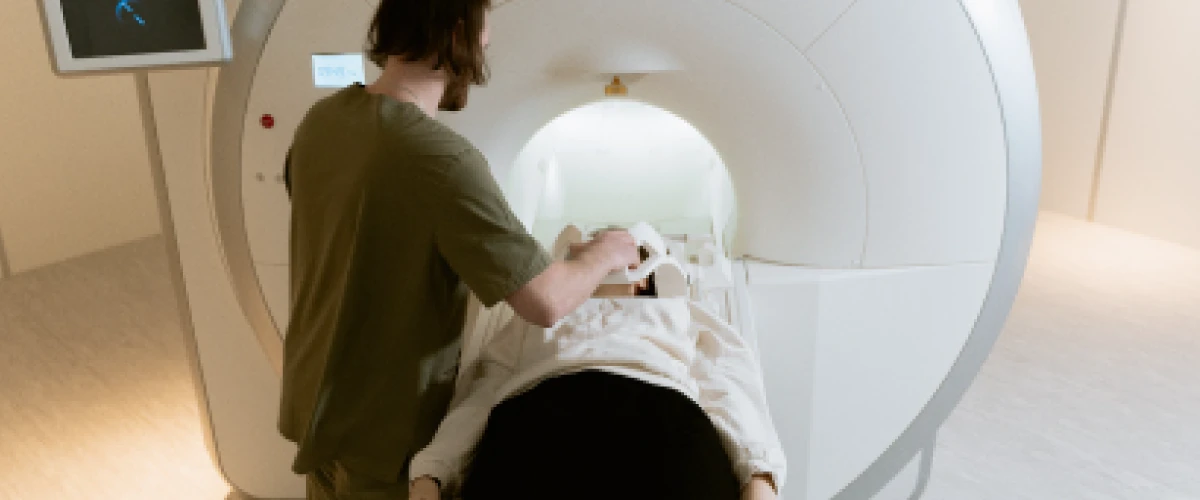
3.Scanning Procedure
- The patient lies on a motorized table.
- The scanner captures both PET (metabolic) and CT (anatomical) images.
- The scan takes approximately 30–60 minutes.
4.Image Fusion & Reporting
- Advanced software combines both images.
- A nuclear medicine specialist and radiologist interpret the results.
The result is a high-resolution 3D image that shows both tumor location and biological activity.
What to Expect Before a PET/CT Scan?
- Fasting for 4–6 hours before the scan.
- Avoid strenuous exercise 24 hours prior.
- Diabetic patients may receive special instructions.
- Inform your doctor if pregnant or breastfeeding.
The procedure is non-invasive and painless.
What Are the Benefits of a PET/CT Scan?
- Early cancer detection
- Accurate staging and metastasis evaluation
- Personalized cancer treatment planning
- Real-time treatment response monitoring
- Detection of recurrence
- Whole-body imaging in a single session
Compared to standalone CT or MRI, PET/CT provides functional + structural imaging, improving diagnostic accuracy in oncology care.
Are There Any Risks or Side Effects?
PET/CT scans are generally safe.
- Radiation exposure is low and within medical safety limits.
- The radioactive tracer is short-lived and eliminated naturally.
- Rare mild allergic reactions may occur.
- Temporary discomfort from lying still.
Your healthcare provider evaluates the benefits versus minimal risks before recommending the test.
Advanced PET/CT Imaging at MGM Cancer Institute
Our One-Stop PET/CT Services Include:
- Expert scan interpretation by experienced radiologists and nuclear medicine specialists
- Image-guided biopsy for accurate cancer diagnosis
- Multidisciplinary tumor board review
- Personalized cancer treatment planning
- Radiation therapy planning support
Our advanced oncology imaging ensures accurate diagnosis, precise staging, and evidence-based treatment strategies.
Conclusion
A PET/CT scan is an essential tool in modern cancer care. By combining metabolic and anatomical imaging, it improves early detection, staging accuracy, and treatment monitoring. For patients undergoing cancer evaluation, PET/CT plays a vital role in guiding personalized and effective treatment.
To consult our Nuclear Medicine experts or schedule a scan, contact MGM Cancer Institute at +91 44 4251 5151.
Frequently Asked Questions
PET/CT scans are highly sensitive in detecting metabolically active cancer cells and are more accurate than standalone CT or MRI for identifying metastasis.
No, the procedure is non-invasive and painless. Patients may only feel mild discomfort during the tracer injection.
Yes, but blood sugar levels must be controlled before the scan for accurate imaging results.
A PET-CT scan usually costs between ₹10,000 and ₹40,000. The price varies depending on the hospital, scan type, and technology used.
Contact Us
Visiting Hours
OPEN 24 hours 7 days a week.
OPD Timings : Monday to Saturday
( 9:00 AM to 5:30 PM )
Appointments
Emergency
Visit the hospital
MGM Cancer Institute
No 119 & 121, Nelson Manickam Road, Raajeswari Street, Rajaram Mehta Nagar,
Aminjikarai, Chennai – 600029


At MGM Cancer Institute, we believe in curing the fear of cancer first. We understand that battling cancer is not just a physical fight, but a mental and emotional one as well. Our dedicated team is committed to providing exceptional healthcare that improves your overall well-being and eases the anxiety that comes with cancer. With a 150-bed facility in the heart of your city, we are here to support you every step of the way.
© MGM Cancer Institute. All Rights Reserved. Last updated on 20-01-2025.